


Occipital nerve pain may look like migraines and tension headaches, therefore, it is sometimes misdiagnosed. Many people with pain at the back of the head, a sensitive scalp, or spreading discomfort are unaware that nerve compression may be the underlying cause.
It is crucial to grasp the difference between dealing with symptoms and seeking long term fixes. We provide patients with comprehensive information about occipital nerve decompression, including the steps involved, the procedure mechanism and its potential role in a broader treatment plan.
Overview of the Occipital Nerves
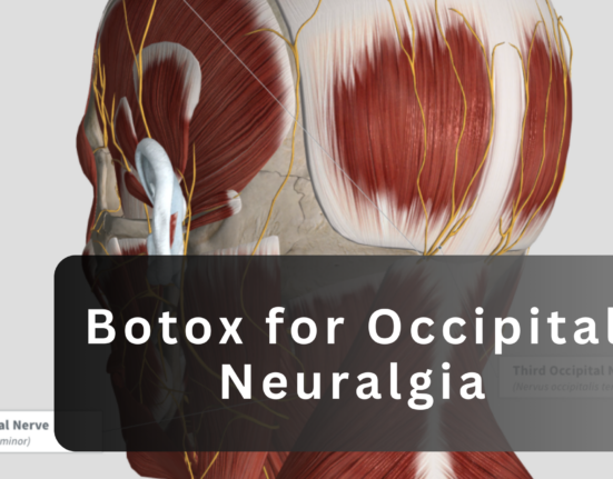
The occipital nerves are located at the back of the head and play a key role in sensation across the scalp. The greater and lesser occipital nerves travel from the upper neck toward the scalp, passing through muscles and connective tissue along the way.
When these nerves become compressed or irritated they can trigger chronic head and neck pain. This compression may be caused by tight muscles, scar tissue, blood vessels or anatomical variations that place pressure on the nerve.
When Occipital Nerve Decompression Is Considered
The occipital nerve decompression is not usually a first line treatment. While typically considered for patients who continue to experience significant symptoms despite conservative care.
Patients may have tried medications, physical therapy, lifestyle modifications, or injections without success. Repeated flare ups and consistent pain patterns often warrant additional investigation.
An Occipital nerve block can aid decision making. A nerve block may temporarily relieve discomfort, suggesting that nerve compression is the cause and making surgical assessment more relevant.
Diagnostic Process Before Surgery
Before surgery is considered a detailed diagnostic process is essential. This usually begins with a thorough clinical examination and review of pain history. Providers assess pain location, triggers, duration and response to previous treatments.
The occipital nerve block for migraine is often used as a diagnostic tool. If the block provides temporary relief and it supports the idea that the occipital nerve is involved. Some patients may experience worse pain after an occipital nerve block which can indicate nerve sensitivity or inflammation and may influence treatment planning.
Preparing for Occipital Nerve Decompression
The preparation for occipital nerve decompression includes a preoperative evaluation to ensure the patient is medically fit for surgery. This may involve routine blood work, imaging review and assessment of other health conditions.
Medication adjustments are often discussed before the procedure. Certain medications may need to be paused or adjusted to reduce surgical risk and support healing.
Step by Step Overview of the Occipital Nerve Decompression Procedure
The technique is typically performed under anesthesia to ensure patient comfort. The patient is positioned to allow safe access to the occipital nerve region.
A careful incision is made in the back of the head or upper neck, depending on the nerve being addressed. The surgeon then identifies the occipital nerve and surrounding structures.
The compression points are carefully examined. These may include tight muscle fibers, connective tissue bands or nearby blood vessels. The surgeon releases or removes tissue that is placing pressure on the nerve.
How the Procedure Relieves Pain
The occipital nerve decompression works by reducing mechanical pressure on the nerve. Once the nerve is no longer compressed, inflammation may decrease, allowing more normal nerve signaling.
This reduction in irritation can lead to fewer pain signals being transmitted to the brain. Over time, patients may experience a decrease in headache frequency and intensity.
Recovery and Post Procedure Care

While immediately after the surgery, patients may experience mild swelling, tenderness or stiffness in the treated area. The symptoms are usually temporary and managed with prescribed pain relief measures.
The early recovery focuses on wound care and avoiding activities that strain the neck or scalp. The patients are given clear instructions on keeping the incision clean and monitoring for signs of infection.
Most patients return to normal daily activities gradually. Full recovery timelines vary, but many individuals resume routine activities within weeks, depending on healing progress.
Risks, Limitations and Expected Outcomes
As with any surgical procedure, occipital nerve decompression carries some risks. Short term side effects may include swelling, numbness or temporary discomfort.
Success rates can vary based on individual anatomy, pain patterns and overall health. Not every patient experiences complete symptom resolution.
While appropriate patient selection is critical and the evaluation helps ensure that surgery is recommended only when it is likely to provide meaningful benefit.
Occipital Nerve Decompression vs Non Surgical Options
Non surgical options such as medications and repeated nerve blocks continue to play an important role in treatment. These approaches may be effective for some patients, especially early in the condition.
Usually, surgery is only contemplated after conservative measures have failed. As part of making an informed choice, readers may find it helpful to read Migraine Surgery Debates, Treatments and Treatments 2025 for a more comprehensive examination of how surgical treatments fit into migraine therapy.
Importance of Specialist Evaluation and Follow Up
Minimally invasive nerve and muscle sparing surgical techniques for occipital neuralgia require careful evaluation by experienced specialists who understand nerve related head pain. Achieving effective results depends on making an accurate diagnosis and selecting the most appropriate surgical approach while follow up visits allow clinicians to monitor recovery that manage symptoms and address concerns early. Long term care plans may also include lifestyle adjustments or supportive therapies to help maintain surgical outcomes.
Conclusion
Occipital nerve decompression is a planned surgery that helps reduce nerve compression that causes long term pain in the head and neck. The patients can make better selections when they know the processes of the operation, how long it will take to recover and what the realistic results are. Individuals can ascertain the suitability of this therapy for their long term treatment objectives by consulting with experienced specialists regarding all available possibilities.